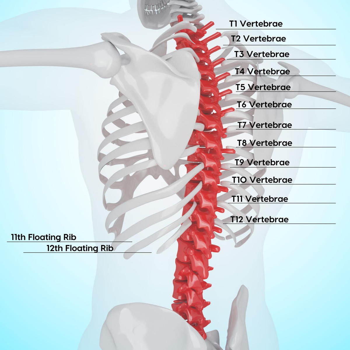
Typical thoracic vertebrae, Radiology Reference Article
Given the twelve thoracic vertebrae are largely similar, most are considered typical thoracic vertebrae with the exceptions T1 and T9 to T12. For a basic anatomic description of the structure of ty

X-ray images of the standard lateral (A) and the coned lateral (B)

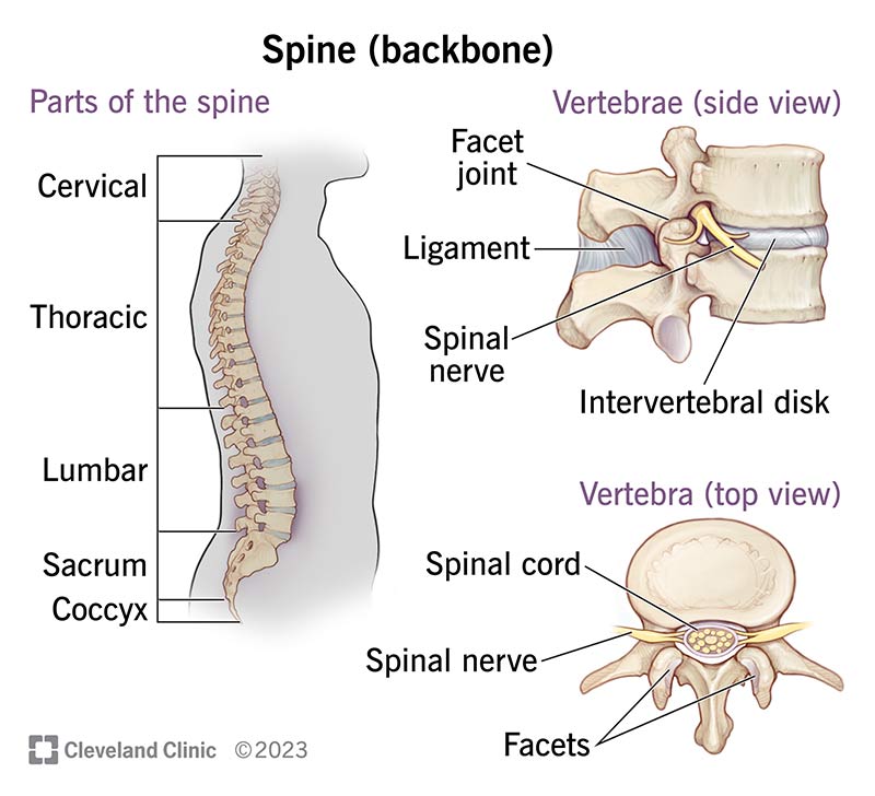
Vertebra, Radiology Reference Article
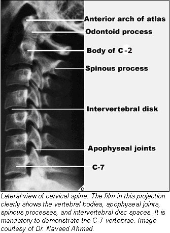
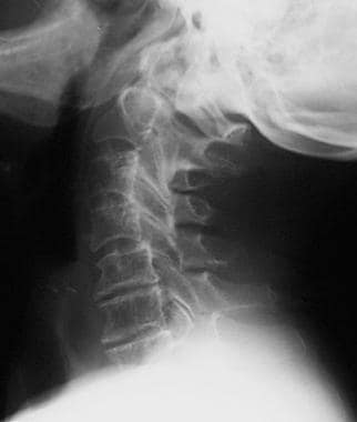
Radiographic positioning techniques for the cervical spine
Rheumatoid Arthritis Spine Imaging: Practice Essentials, Radiography, Magnetic Resonance Imaging

Syndesmophyte, Radiology Reference Article

Ventrodorsal projection radiograph of the thoracic vertebral column.

Cervical and Thoracic Spine: Normal Variants and Artifacts

Pediatric Vertebral Body Anomalies, Pediatric Radiology Reference Article, Pediatric Imaging
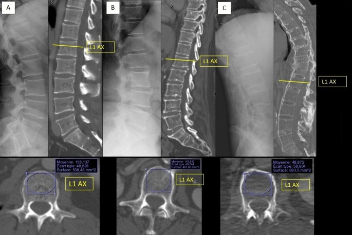
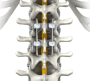
Relationship between spinal structural damage on radiography and bone fragility on CT in ankylosing spondylitis patients

Cervical spine alignment, Radiology Reference Article