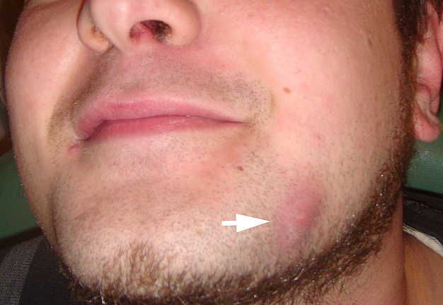
a Mandibular fistula indicated by an arrow in the apical region of dd
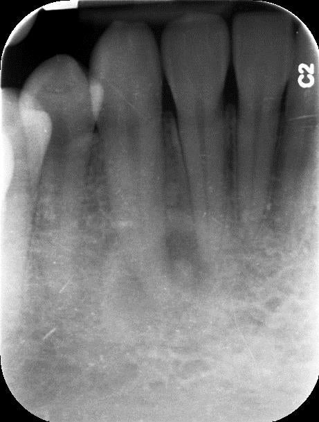
Download scientific diagram | a Mandibular fistula indicated by an arrow in the apical region of dd 36-37. b A fistula in the apical region of dd 46-47 (white arrows) and a red area in the mucosa (black arrows) are seen in the right lingual surface of the mandible. c Panoramic radiograph showing no bone lesions in the mandible. d Periapical x-ray with no bone involvement in the apical region of dd 46-47 from publication: Treatment of bisphosphonate-induced osteonecrosis of the jaws with Nd:YAG laser biostimulation | Osteonecrosis, Jaw and Nd:YAG Laser | ResearchGate, the professional network for scientists.

SciELO - Brazil - Differential diagnosis and clinical management of periapical radiopaque/hyperdense jaw lesions Differential diagnosis and clinical management of periapical radiopaque/hyperdense jaw lesions

Medication-related osteonecrosis of the jaw without osteolysis on computed tomography: a retrospective and observational study
Single and Multiple Odontogenic Cutaneous Sinus Tracts

a Mandibular fistula indicated by an arrow in the apical region of dd
Cureus, Cemento-Osseous Dysplasia: A Detailed Comparison of the 2005 and 2017 WHO Classifications and Case Analysis

World Small Animal Veterinary Association Global Dental Guidelines - Niemiec - 2020 - Journal of Small Animal Practice - Wiley Online Library
Frs hfn

Satu ALALUUSUA, University of Helsinki, Helsinki, HY, Institute of Dentistry

a Mandibular fistula indicated by an arrow in the apical region of dd
Case Archive, School of Dental Medicine

Dental CT: Pathologic Findings in the Teeth and Jaws

Malformations of the tooth root in humans. - Abstract - Europe PMC