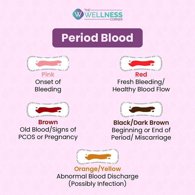
Red Cell Staining (Color) • The Blood Project
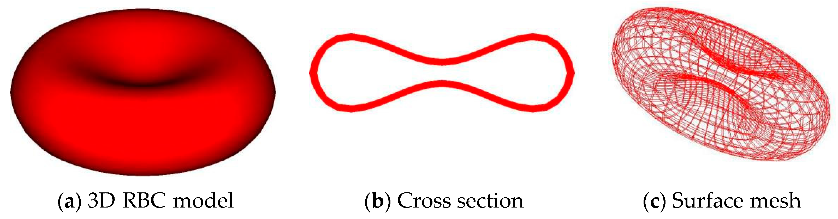
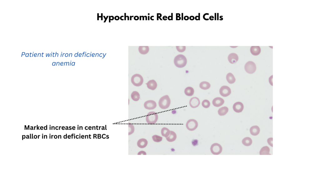
Central pallor Introduction A normal red blood cell has a biconcave disk shape. Because the center is much thinner than the periphery, it creates the
Red Cell Staining (Color) • The Blood Project

4,900+ Red Blood Cell Microscope Stock Photos, Pictures & Royalty

Human Blood Film Slide, Smear, Wright's Stain: Prepared Microscope

Immunofluorescence staining protocol for STED nanoscopy of
Behind the Scenes of Visible Biology: Bringing the Blood Cell Lab

Staining in Microbiology Meaning, Types & Techniques - Lesson

Blood: The Good, the Bad, and the Ugly – Microbiology: A

H&E stain - Wikipedia
Cell Tracking Red Dye Kit - Longer cell staining, DMSO-free

Immunofluorescence staining protocol for STED nanoscopy of

Blood Smear Test, Procedure & Possible Results - Lesson