Pitfalls of inferior vena cava M-mode – NephroPOCUS
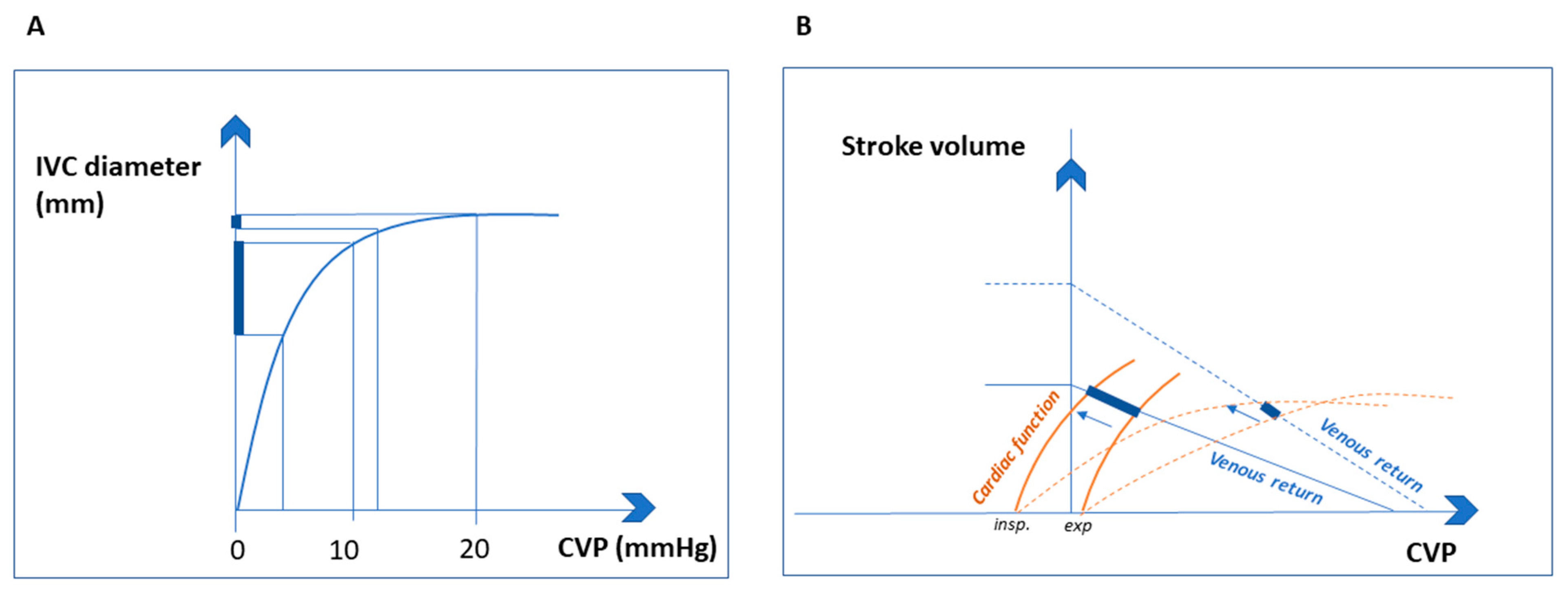
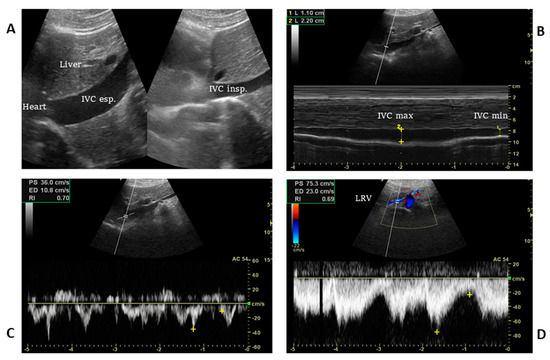
Visual estimation of IVC collapse on B-mode (grey scale image) is generally preferred to M-mode, though in theory, M-mode measurement might be able to give accurate collapsibility index. There are several reasons for this. A major limitation of IVC M-mode is that the vessel moves mediolaterally and craniocaudally during respiration, with collapse occurring off axis…

Cardiac – Page 2 – NephroPOCUS

PDF) Transcending boundaries: Unleashing the potential of multi-organ point-of-care ultrasound in acute kidney injury
Inferior Vena Cava POCUS: The Basics of Image Acquisition - Renal

Venous Excess Doppler Ultrasound for the Nephrologist: Pearls and Pitfalls - ScienceDirect
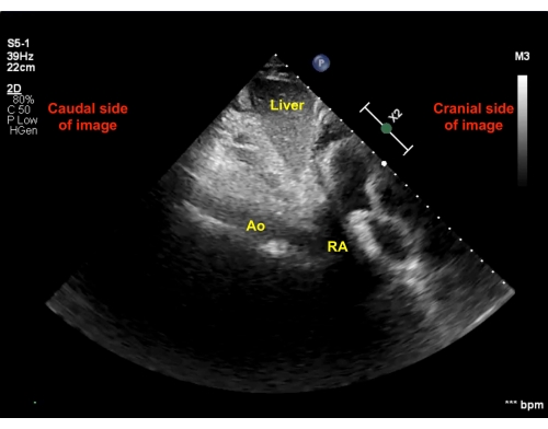
Image Acquisition Method for the Sonographic Assessment of the

Cardiac – Page 3 – NephroPOCUS

The 'ring of fire' Foley balloon – NephroPOCUS
JCM, Free Full-Text
Transcending boundaries: Unleashing the potential of multi-organ
Abhilash Koratala – Page 9 – NephroPOCUS

The 'ring of fire' Foley balloon – NephroPOCUS

Changes in the Inferior Vena Cava Are More Sensitive Than Venous
JCM, Free Full-Text

Subcostal longitudinal view of the inferior vena cava with

PDF) Transcending boundaries: Unleashing the potential of multi