Pelvic floor parameters predict postpartum stress urinary incontinence: a prospective MRI study, Insights into Imaging
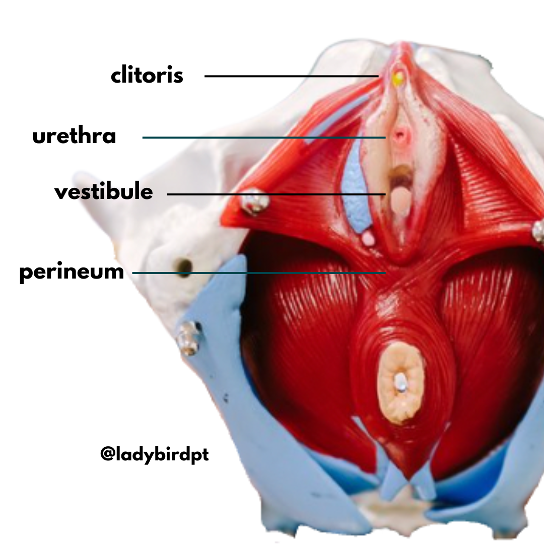
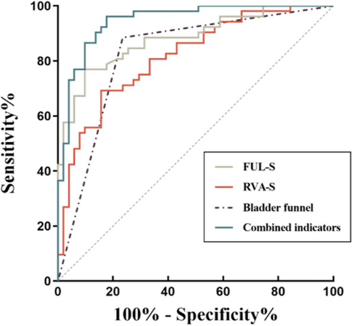
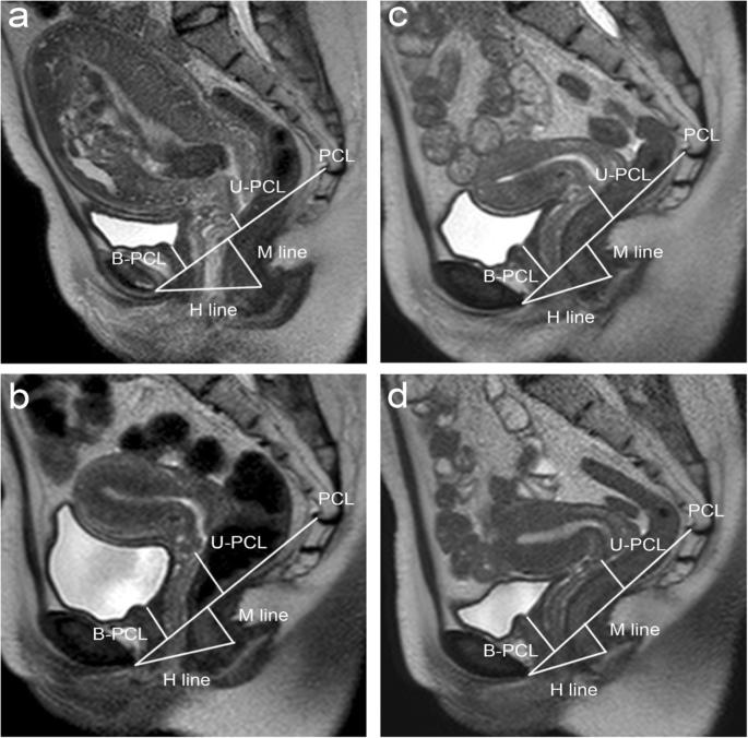
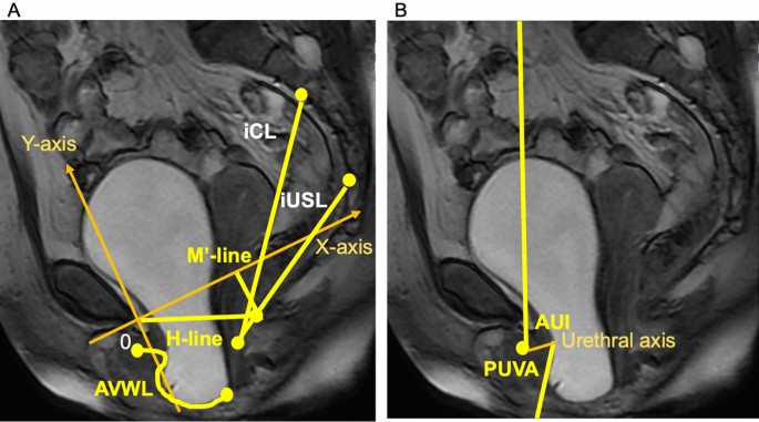
Objective To investigate the pelvic floor changes in primiparas with postpartum stress urinary incontinence (SUI) after vaginal delivery using pelvic floor MRI. Materials and methods Fifty-two women were enrolled in the primiparous stress urinary incontinent (PSUI) group and 51 in the primiparous continent (PC) group. Thirty nulliparas were also recruited as the nulliparous control (NC) group. Levator ani muscle (LAM) injury, levator hiatus area (LHA), H-line, M-line, the distance from the bladder neck and cervix to the pubococcygeal line (B-PCL and U-PCL), levator plate angle, the anterior angle of the urethra, bladder neck descent, retrovesicourethral angle, functional urethral length, and a bladder neck funnel were evaluated on MRI images. Univariate and multivariate logistic regression analyses were used to explore anatomical predictors for SUI. Results The primiparas in the PSUI group showed more obvious LAM injuries than in the PC groups (p = 0.001). LAM function assessment: the PSUI group had larger LHA and shorter B-PCL and U-PCL than the other groups during straining. Assessment of urethral mobility and function: the PSUI group had larger anterior angle of the urethra, bladder neck descent, retrovesicourethral angle, and shorter functional urethral length than the other two groups (all p < 0.05). Up to 88.5% of primiparas in the PSUI group showed bladder funnel (p < 0.001). The logistic regression analysis showed that retrovesicourethral angle, functional urethral length, and the presence of bladder funnel were significantly associated with postpartum SUI (p < 0.05). Conclusions Increased retrovesicourethral angle, shortened functional urethral length, and the presence of bladder funnel may be anatomical predictors for SUI in the early postpartum period. Urethral sphincter dysfunction plays an essential role in developing postpartum SUI. Critical relevance statement This study used several measurements to reflect the anatomical structure and functional changes of the pelvic floor to identify the best anatomical predictors associated with postpartum stress urinary incontinence (SUI), aiming to provide new insights into treatment strategies for postpartum SUI. Key points • Increased retrovesicourethral angle, shortened functional urethral length, and the presence of bladder funnel are more commonly seen in primiparas with SUI. • The combination of retrovesicourethral angle, functional urethral length, and bladder funnel had the highest diagnostic performance in predicting postpartum SUI (AUC=0.947). • Urethral sphincter dysfunction may be the main pathophysiological foundation in SUI development. Graphical Abstract

Risk factors for postpartum stress urinary incontinence: a

PDF) Stress urinary incontinence: pre-pregnancy history and effects of mode of delivery on its postpartum persistency
Cureus The Effectiveness of Pelvic Floor Muscle Exercise in
Pelvic floor assessment using magnetic resonance imaging after vaginal delivery and elective caesarean delivery

IJERPH, Free Full-Text

above): shows a number of possible measurements using MRI imaging
MRI of the pelvic floor in female patients with stress urinary
Pelvic floor parameters predict postpartum stress urinary

Evaluation of pelvic structural abnormalities in primiparous women

PDF) The pathophysiology of stress urinary incontinence: a
Association between overactive bladder and pelvic organ mobility

A model identifying characteristics predictive of successful

Pregnancy-induced remodeling of the murine reproductive tract: a