Large pedunculated polyp left breast nipple.

Diagnostic Endoscopy
T1-weighted MRI: Tumor of the posterior cranial fossa (single arrow) as

Magnetic resonance imaging showing a large well-defined cystic lesion
Atlas of breast cancer early detection

Thorax CT scan (A,B) is showing two nodules of the right upper and
A clinical case of large fibroepithelial polyp of breast nipple - International Journal of Case Reports and Images (IJCRI)
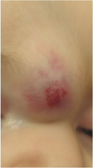
Frontiers Childhood Vascular Tumors

Leiomioma de mama. Imagen histológica 20x.

Thorax CT scan (A,B) is showing two nodules of the right upper and

Paget's Disease of Breast (PDB)

Cutaneous Disorders of the Breast

PDF] A clinical case of large fibroepithelial polyp of breast nipple

A clinical case of large fibroepithelial polyp of breast nipple - International Journal of Case Reports and Images (IJCRI)

Contrast-enhanced CT scan in axial views of neck. A 36 × 48 mm soft
T1-weighted MRI: Tumor of the posterior cranial fossa (single arrow) as