Optical Coherence Tomography: Imaging Mouse Retinal Ganglion Cells In Vivo
Scientific Article | Structural changes in the retina are common manifestations of ophthalmic diseases.

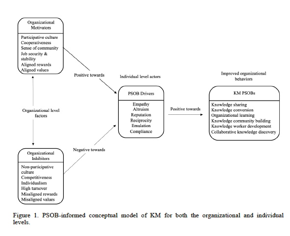
Multimodal Coherent Imaging of Retinal Biomarkers of Alzheimer's Disease in a Mouse Model

Retinal neurovascular responses to transcorneal electrical stimulation measured with optical coherence tomography - Xiaofan Su, Hao Zheng, Qian Li, Pengcheng Sun, Meixuan Zhou, Heng Li, Jiahui Guo, Xinyu Chai, Chuanqing Zhou, 2020

Topical Nerve Growth Factor (NGF) restores electrophysiological alterations in the Ins2Akita mouse model of diabetic retinopathy - ScienceDirect

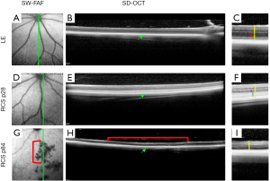
Adaptive optics with combined optical coherence tomography and
In vivo retinal imaging in translational regenerative research - Sher - Annals of Translational Medicine

All Protocols and Video Articles in JoVE
Methods paper on in-vivo cellular resolution neuronal and vascular retinal imaging published - Burns & Pugh Lab

Researching Review of Advances in Ophthalmic Optical Imaging Technologies from Several Mouse Retinal Imaging Methods
Application of Optical Coherence Tomography to a Mouse Model of Retinopathy

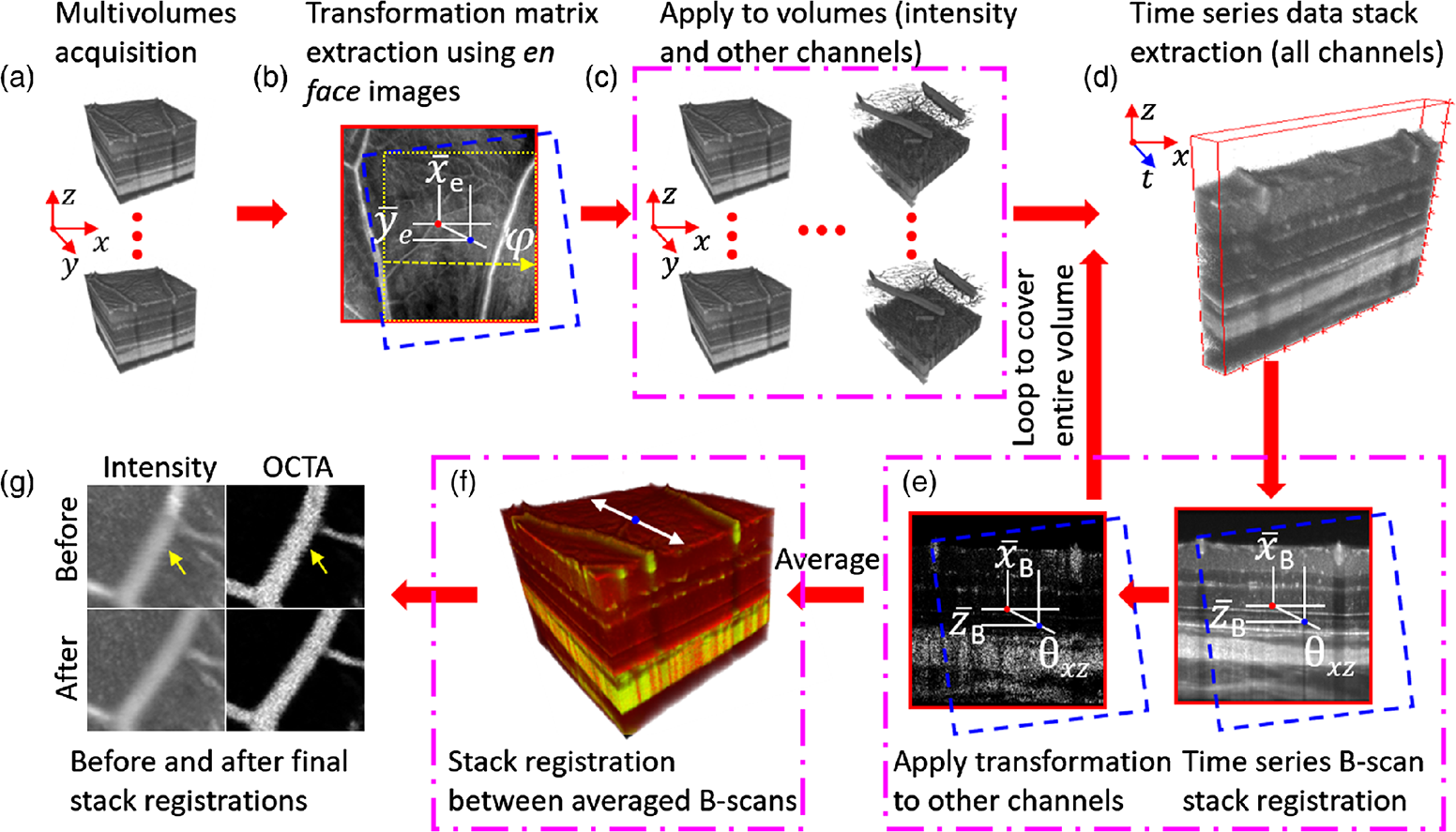
Optical coherence tomography angiography (OCT-A) in an animal

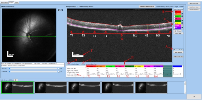
Image segmentation of mouse eye in vivo with optical coherence